
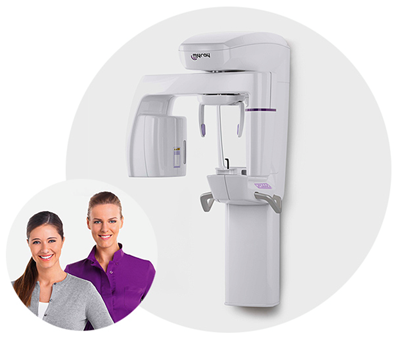
The present and the future of my work. In three dimensions. Hyperion X9 offers me multiple possibilities and a full range of functions to aim for the best, at all times. MyRay, Just right for you.

All the possible choices in one touch. Hyperion X9 adapts to your work, optimizes your time, satisfies your needs. A hybrid platform with exceptional performances. Hyperion X9 unites advanced technology and simplicity of use, thereby ensuring you excellent diagnostic analyses in a short time. Try out a new level of efficiency thanks to the automatic alignment of the 2D and 3D detectors. Adapt the platform to your needs: three exceptional solutions, easy to adapt and flexible. Hyperion X9 offers you a wide range of 2D analyses, cephalometric projections and all the best of 3D technology.
Hyperion X9 adopts a FOV up to 11 cm of diameter, in order to allow us complete diagnoses with maximum efficiency.
You will thus have at your disposal the whole dentition, including the complete roots of the third molars (the wisdom teeth) and the surrounding bone structures. No limit to the planning of multiple implants, even with the use of surgical guides.
Volumetric acquisition with a full 360° scan capable of eliminating the artefacts of the resulting image. High resolution at extremely low X-ray doses: excellent quality, detailed particulars, fast diagnosis. Hyperion X9 ensures you excellence with a maximum voxel resolution of 75 μm across the full arch. The constant potential generator with pulsed mode emission automatically optimizes the parameters according to the patient, thereby ensuring maximum results with minimum exposure (3.6 s).
Stability and comfort: choose the best with as many as 7 support points for the sake of a stable patient under all circumstances. With a seat for the chin, the adjustable self-locking forehead support and the replaceable bite block, the positioning system has never been so simple and effective. Perfect images in every situation (3 patent pending).
Hyperion X9 offers an efficient dedicated detector for 2D diagnostics (PAN/CEPH), relocatable and releasable, fastened onto a safety device; with the possibility of adding a second detector so as to carry out cephalometric projections.
Hyperion X9 offers you the best of 2D programs, from the panoramic exams to the cephalometric ones, with a rapid exposure so as to contain the times and reduce the X-rays dose for the patient’s safety.
Hyperion X9 brings you maximum speed in image sharing. Simple, practical, effective. Under all circumstances.
Hyperion X9 facilitates your work through the innovative patented MRT (Morphology Recognition Technology). Automatically secure the recognition of the patient’s morphology for a correct X-ray exposure and an optimal image. With the aid of MRT, there is no need to plan exposure times or technical factors such as the kV or mA level. Hyperion X9 avoids over- or under-exposed images, thereby preserving the quality of your diagnostics and avoiding useless radiogenic doses to the patient (2 Patent Pending).
| 3D IMAGES | EXTENDED FOV | FULL FOV |
|---|---|---|
| Detector technology | Amorphous Silicon – Csi (Cesium Iodide) Scintillator | Amorphous Silicon – Csi (Cesium Iodide) Scintillator |
| Dynamic range | 16 bit (65535 grey shades) | 16 bit (65535 grey shades) |
| Minimum scan time | 18 s | 18 s |
| Rotation | 360° | 360° |
| Image voxel size | 75 μm (minimum slice thickness) | 75 μm (minimum slice thickness) |
| Field of view, diameter x height | 108x80 mm | 108x50 mm |
| Available FOV sizes (Øxh) | 11x13e - 11x8 - 8x8 - 11x5 - 8x5 - 5x5 cm | 11x8e - 11x5 - 8x5 - 5x5cm |
| Largest size of image dataset | 720 MB | 450 MB |
| X-ray exposure time | 3.6 s (High Resolution) - 9.0 s (Peak Resolution) | 3.6 s (High Resolution) - 9.0 s (Peak Resolution) |
| Typical effective dose (ICRP 103): 11x8 FOV | 33.5 μSv (High Resolution) – 78.6 μSv (Peak Resolution) | 33.5 μSv (High Resolution) – 78.6 μSv (Peak Resolution) |
| Patient Alignment | Servo-assisted: “Scout View” method | Servo-assisted: “Scout View” method |
| Image format | Exclusive iRYS and DICOM 3.0 software | Exclusive iRYS and DICOM 3.0 software |
| Minimum render times for CB3D data | 15 s | 15 s |
Compact and easy to use. Capable of being installed onto any wall suitable for an intraoral X-Ray. Out of our experience, this product represents the best solution for every dentist.
A refined, clean, essential, ultra-compact design. Hyperion X5 is the smallest in the world and can be installed in any practice: all you need is one wall!
Light and compact like an intraoral X-Ray, for a full world of possibilities.All you need is one wall.
The diagnostic features you need in just one touch. Accurate diagnoses for different requirements, from adults to children, essential programs for your needs, in a few, simple steps.
User-friendly. Quick. Setting-free. Hyperion X5 gathers in itself all the technological innovations that simplify the workflow of your practice. Everything proves therefore to be simple and intuitive. Immediate access, facilitated visual control, intuitive interface. Interacting with a panoramic imager has never been so simple.
Hyperion X5 shares its own images with all the PC’s present in the practice thanks to the image management iRYS software, common to every MyRay device. It is possible to send images with a TWAIN protocol to every compatible management software. Hyperion X5 is an open system: the virtual console, available both for WINDOWS systems and as iPad APP*, enables a remote control of the machine and a visualization of the images acquired on tablet. Managing and archiving the images is practical and quick: because of that, we have conceived a powerful platform capable of interfacing with third party systems, thanks to the DICOM protocols and other communication methods
| IMAGES 2D | |
| Type | Adult and child panoramic*, QuickPAN, MultiPAN, “Bitewing”* dentition, PA and LL (right and left) maxillary sinuses, Temporomandibular Joint (2 x LL +2 x PA) open and closed mouth. |
|---|---|
| Child examination | Yes |
| Maximum resolution | from 5 to 7 lp/mm |
| Maximum field of view (mm) | 280 (length); 150 (height) |
| Reduced fields of view (cm) | 6 x 12.5* (Child) 6 x 9* (Dentition bitewing) |
| Maximum image dimensions | 7.5 MB |
| Magnification | PAN: 1.2 - 1.3 |
| Scan time | PAN 12s (STD.) – 6.6s (Quick Scan) |
| Typical effective dose (ICRP 103) | PAN: 5 - 9 μSv |
| Minimum image display times | RealTime |
| Advanced filters | PiE (Panoramic image Enancher) |
| CONNECTIVITY | |
| Connections | LAN / Ethernet |
| Software | iRYS |
| Supported protocols | DICOM 3.0, TWAIN, VDDS |
| DICOM nodes | IHE certification (Print; Storage Commitment; WorkList MPPS; Query Retrieve) |
| App | Compatibility with iPad and iPhone |

Hyperion X9 pro offers the very best of 3D technology, cephalometric projections and a wide range of 2D examinations.
Maximum flexibility for your diagnoses. Hyperion X9 pro is fully configurable and its modular and scalable design makes it possible to transition from a basic to a more advanced version in an easy and cost-effective manner. An extraordinary platform that adapts to the needs of your dental practice thanks to the 2D PAN/CEPH sensor that can be easily relocated and to the reversible teleradiographic arm which can be installed on both sides. The most compact 3-in-1 hybrid system available on the market for high-quality 2D and 3D examinations.
High-definition images, extremely sharp details, upgraded MultiPAN system for maximum results in every situation.
MultiFOV and high resolution: wonderfull 3D images for all your radiology needs.
Hyperion X9 pro optimises your work, adapts to your needs and allows to focus on what’s really important: your diagnoses.
Hyperion X9 pro is equipped with a user-friendly interface, also available in the iPad-specific application, for an easy and intuitive control. In few simple steps you can choose and set up the most appropriate exam based on the clinical and anatomical relevance.
The multi-platform control panel gives you easy and immediate access to all the device features. The interface guides you step by step, from the exam selection to its preparation, with FOV guided positioning. The result is easier, faster and more effective examinations.
User-friendly graphics and direct controls make your work easier, providing patients with a more relaxing experience. Hyperion X9 pro is characterised by the simplicity of use and the rapidity of procedures, such as the possibility to choose predetermined programmes directly from the homepage. The control panel interface provides precise instructions on the patient’s positioning depending on the selected protocol.
The positioning is fast and accurate thanks to an alignment system with 4 laser beams projected directly on the patient’s face and to the state-of-theart ergonomic head support unit equipped with 7 fixing points for maximum stability during scanning. The Face to Face positioning guarantees maximum freedom of movement and the patient’s comfort.
Through the Scout View system it is possible to centre the volume on the area of interest, while the patient can remain in the same comfortable position. From the PC, the operator can view the two images (sagittal and frontal) at very low irradiation and accurately modify the scanning area letting the equipment, supplied with servo-assisted movements, find the correct position. This procedure eliminates the risk of having to repeat the examination.
Thanks to advanced QuickScan protocols, available for both 2D examinations and 3D cquisitions, it is possible to obtain accurate images with lower doses as compared to a standard acquisition. These protocols are the ideal tool for post-surgery check-ups and for the identification of any macrostructures (such as impacted teeth or dental agenesis).
The best all-in-one software platform for 2D and 3D
| 3D IMAGES | FOV 10x8 VERSION | FOV 13x16 VERSION |
|---|---|---|
| Detector technology | Amorphous silicon - CsI with direct deposition | Amorphous silicon - CsI with direct deposition |
| Dynamic range | 16 bit (65535 livelli di grigio) | 16 bit (65535 grey levels) |
| Typical scan time | 14,4 s | 14,4 s |
| Rotation | 360°/180° | 360°/180° |
| Image voxel size | Minimum 75 μm | Minimum 68 μm |
| Field of view, diameter x height | 108 x 80 mm | 108 x 50 mm |
| Available FOV sizes (Øxh) | 6x6 - 8x6 - 8x8 - 10x6 - 10x8 | 6x6 - 8x6 - 8x8 - 10x6 - 10x8 10x10 - 13x8 - 13x10 - 13x16 4x4 - 7x6 (eXtended Functionality) |
| Typical image size | 495 MB | 820 MB |
| Minimum scan time | 6,4 s | 3,6 s |
| Typical X-ray exposure time | 1.6 s (Low-dose QuikScan) - 8.0 s (SuperHD Mode) | 1.6 s (Low-dose QuikScan) - 8.0 s (SuperHD Mode) |
| Patient alignment | Servo-assisted: Scout View method | Servo-assisted: Scout View method |
| Image format | Exclusive iRYS and DICOM 3.0 software | Exclusive iRYS and DICOM 3.0 software |
| Minimum render times for CB3D data | 15 s on average | On average, real-time for FOV XF 4x4 QuickScan |
| CONNECTIVITY | |
| Connections | LAN / Ethernet |
| Software | MyRay iRYS and App iPad |
| Supported protocols | DICOM 3.0, TWAIN, VDDS |
| DICOM nodes | IHE- compliant (Print; Storage Commitment; WorkList MPPS; Query Retrieve) |
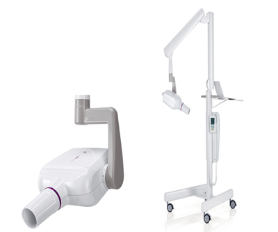
RXDC - High frequency intraoral x-ray unit
Highest quality with lowest exposure.
Always-sharp images, versatility and meticulous attention to patient health.
With RXDC you get the best DC technology with the lowest X-ray dose.
The constant potential high frequency generator (DC) provides sharp images with the very highest level of detail.
Compared to AC systems, they also reduce exposure times and the amount of harmful radiation by containing the dose administered to the patient.
A focal spot of just 0.4 mm - one of the smallest available - ensures images are always sharp and of the highest quality. High definition real-time imaging.
Superb image definition: sharp edges and excellent detail.
An embedded collimator cone gives a source-to-skin distance of 30 cm.
This increases X-ray parallelism, providing more precise images, lower doses and ensuring greater attention to patient health.
RXDC offers maximum flexibility and optimum X-ray quality whatever the type of sensor connected.
Multi-Mode automatic exposure parameter modulation always ensures optimal time and power selection. Parameters are, in fact, adjusted automatically according to patient build and the region under investigation.
Attention to patient health is meticulous thanks to the constant potential DC generator with adjustable power (from 8 to 4 mA). Moreover, rectangular collimators can be used: these reduce the irradiated body area and thus lower the dose received by the patient.
The ergonomic handle is designed to maximise grip comfort and ensure easy, stable positioning of arms and tube head. A protractor with a graduated scale allows optimal repositioning of the tube head.
Simple installation, versatility, reliability RXDC provides outstanding adaptability and simplicity of installation thanks to extruded aluminium arms with an integrated self-balancing system that can be pointed in 6 directions - available in lengths of 40 cm, 60 cm and 90 cm. All parts are made from materials of only the finest quality to minimize maintenance costs and reduce the risk of accidental vibration during acquisition.
Wall-mounted with variable positions or in a mobile cart-mounted version (to be shared among multiple workstations), RXDC is extremely versatile and easily adapts to all your working needs.
| TECHNICAL DATA | |
|---|---|
| Generator | Constant potential, microprocessor-controlled |
| Working frequency | 145 - 230 KHz with self-adjustment (typically 175 KHz) |
| Focal spot | 0.4 mm (IEC 336) |
| Total filtration | 2.0 mm Al @ 70 kV |
| Anode current | 4 / 8 mA |
| Voltage at X-ray tube | 60 / 65 / 70 kV (*) |
| Exposure times | 0.020 – 1.000 seconds, R’10 and R’20 scale |
| Source-skin distance | 20 and 30 cm |
| Irradiated field | Ø 55 mm and Ø 60 mm round |
| Additional collimators | 35 x 45 mm rectangular, 31 x 41 mm and 22 x 35 mm, for sensors size 2 and size 1 |
| Power supply | 50/60 Hz, 115-120 V AC ±10% or 230-240 V AC ±10% |
| Duty Cycle | Continuous operation with self-adjustment up to 1s/80s total |
| Arms (for Standard version only) | Available in 3 lengths: 40 cm – 60 cm – 90 cm |
| Max. arm extension | 230 cm, from wall |
| Certification | CE 0051, FDA approved |
| Versions | Standard (wall mounted) or Mobile (on portable cart) |

RXDC - Intraoral x-ray unit with eXTend wireless technology
Maximum image quality, minimum dose for the patient.
RXDC - eXTend technology provides always-sharp images, a full configuration range and the exclusive flexibility of wireless technology.
This technology gives sharp images with greater detail and lower exposure times than would be attainable with AC X-ray units, which are characterised by variable emissions.
Moreover, constant-potential design ensures image generation is unaffected by power fluctuations.
RXDC - eXTend technology is reliable for all diagnostic needs and always provides high-definition images by adapting to the sensor type.
The focal spot of just 0.4 mm is placed in the tube head in such a way as to obtain a source-to-skin gap of 30 cm (total bulk remaining equal).
In this way RXDC - eXTend technology implements extensive internal collimation of the X-rays and gives an extremely small focal spot, producing ever-sharper images and ever-more precise detail.
Automatic parameter modulation ensures the best exposure power/time selection: parameters are automatically determined on the basis of the patient’s build and the specific region of investigation. With 28 selectable sensitivity levels, sharp images are guaranteed with any sensor.
Attention to patient health is meticulous: a high frequency, constant potential generator minimises exposure times and reduces harmful radiation. Where deemed appropriate, the 4 mA mode halves the amount of X-rays. The interchangeable rectangular collimator cone (at 30 cm) further reduces the rradiated body surface area by adapting it to the effective surface area of the sensor.
No downtimes as a result of tube overheating, not even when repeated use is required.The fast dynamic duty cycle allows, in fact, sequential exposures by keeping tube temperature under constant control on the large hand-held unit display.
The efficiency of wireless technology with maximum simplicity of use.
The wireless controller frees users from the limits posed by on-machine control panels or wall-mounted controls. It is equipped with a button for ultra-fast shooting (fraction of a second) and two simple settings which make it easy to select the most suitable X-ray acquisition programme.
The solid extruded aluminium arms are made of high quality materials that ensure strength and durability while reducing the risk of accidental vibration during acquisition.
They are available in lengths of 40 cm, 60 cm and 90 cm and can be pointed in 6 directions to provide maximum adaptability and simplicity of installation.
| TECHNICAL DATA | |
|---|---|
| Generator | Constant potential, microprocessor-controlled |
| Working frequency | 145 - 230 KHz with self-adjustment (typically 175 KHz) |
| Focal spot | 0.4 mm (IEC 336) |
| Total filtration | 2.0 mm Al @ 70 kV |
| Anode current | 4 / 8 mA |
| Voltage at X-ray tube | 60 / 65 / 70 kV (*) |
| Exposure times | 0.020 – 1.000 seconds, R’10 and R’20 scale |
| Source-skin distance | 20 and 30 cm |
| Irradiated field | Ø 55 mm and Ø 60 mm round |
| Additional collimators | 35 x 45 mm rectangular, 31 x 41 mm and 22 x 35 mm, for sensors size 2 and size 1 |
| Power supply | 50/60 Hz, 115-120 V AC ±10% or 230-240 V AC ±10% |
| Duty Cycle | Continuous operation with self-adjustment up to 1s/80s total |
| Arms (for Standard version only) | Available in 3 lengths: 40 cm – 60 cm – 90 cm |
| Max. arm extension | 230 cm, from wall |
| Certification | CE 0051, FDA approved |
| Versions | Standard (wall mounted) or Mobile (on portable cart) |

RXDC, HyperSphere technology
Innovative design, revolutionary ergonomics, advanced technology. RXDC - HyperSphere technology brings the best of DC X-ray units into your surgery.
HyperSphere technology gives the RXDC unit full rotation capability.
The tube revolves freely around the joint, allow it to reach practically any position, including the vertical. RXDC - HyperSphere technology also features an automatic touch-sensitive device for simple, efficient locking/release of the X-ray head tube so it can be repositioned effortlessly between one exposure and the next. Ergonomic zones on the sides of the head provide a firm grip for effective positioning.
The wireless remote controller lets the user control the device (by communicating with the X-ray tube) while enjoying full freedom of movement.
Access to exposure programmes is provided via two simple settings. The large display shows the sequential exposure monitor and the patient exposure dose; moreover, the controller has a wireless X-ray snapshot button. Wireless device control allows fast, easy installation: no fixed control panels are required, thus providing greater freedom when positioning the X-ray unit.
With a tiny focal spot of 0.4 mm (at 30 cm), RXDC - HyperSphere technology produces sharp images under any condition.
The tube head is now even more powerful as it operates at 70 kV, 8 mA. RXDC - HyperSphere technology gives your surgery the precision and quality of cuttingedge know-how.
The constant potential high frequency (DC) generator reduces the most harmful low energy radiation that is characteristic of analogue (AC) generators: current is adjustable (from 8 mA to 4 mA), as are exposure times.
Moreover, the long cone (30 cm) with incorporated rectangular collimator reduces the exposed surface area. This maximises image quality and safeguards patient and worker health.
Maximum flexibility to meet your diagnostic needs. Automatic parameter modulation ensures exposure power and time are always selected according to the patient’s build and the specific region of investigation.
The dynamic service cycle allows uninterrupted use of the RXDC, as in the case of systematic examinations, and real-time monitoring of tube head temperature on the large wireless controller display.
RXDC - HyperSphere technology features a 30 cm cone with a circular and rectangular collimator. The rectangular collimator is ideal for reducing the X-ray dose administered to the patient as it limits the irradiated body area to the effective capture capacity of the sensor.
| TECHNICAL DATA | |
|---|---|
| Generator | Constant potential, microprocessor-controlled |
| Working frequency | 145-230 KHz (typically 175 KHz) |
| Focal spot | 0.4 mm (IEC 336) |
| Anode current | 4 / 8 mA |
| Voltage at X-ray tube | 60 / 65 / 70 kV (*) |
| Exposure times | 0.020 – 1.000 seconds, R’10 and R’20 scale |
| Source-skin distance | 20 e 30 cm |
| Irradiated field | 35 x 45 mm rectangular, Ø 55 mm and Ø 60 mm round |
| Additional collimators | 31 x 41 mm and 22 x 35 mm, for size 2 and size 1 sensors |
| Total filtration | 2.0 mm Al @ 70kV |
| Power supply | 50/60 Hz, 115-120Vac ±10% o 230-240Vac ±10% |
| Duty Cycle | continuous operation with self-adjustment up to 1s/80s total |
| Stability | Automatic lock/release, with touch-sensitive activation (HyperSphere technology) |
| Arms | Available in 3 lengths: 40 cm - 60 cm - 90 cm |
| Max. arm extension | 230 cm, from wall |
| Certification | CE 0051, FDA approved |

Phosphor Plate scanner
High quality digital imaging with the convenience and user-friendliness of traditional film. Hy-Scan phosphor scanner is the perfect balance between technology and tradition.
A compact, fast and easy to use device, producing high resolution intraoral images for consistently reliable diagnostic results.
With its essential and compact design, the Hy-Scan scanner is an ideal tool for any dental surgery.
It is extremely versatile and can be both installed horizontally on a table top, or wall-mounted vertically with a special bracket.
Height definition
High quality images for any application
Hy-Scan is the ideal tool for all clinical applications - endodontics, implants and implant surgery, periodontics, detection of caries - always producing the best high definition images with 34 pixels/mm resolution.
The scanner is compatible with four plate sizes for the capturing of :
images with 30μm pixel size
Acquisition
User-friendliness and maximum workflow efficiency.
Hy-Scan has a servo-assisted system that accepts and scans in a fully automatic (TOUCH-FREE) manner the exposed plates, detects the size, uploads the image to a PC and deletes data from the plate making it ready for the next scan.
Position the plate easily and conveniently to capture high-resolution images. After inserting the plate, the scanner will quickly scan it. The images are then transferred to the PC, viewed and shared with iRYS or another viewing software, printed and sent by e-mail.
Easy connect
Quickly imports data via a USB connection.
Stores and displays the captured images on the PC with iRYS, the all-in-one diagnostic software, with the handy comfortable viewer APP for iPad or any other image management or viewer software with TWAIN interface.
Simplicity and quality
A compact, fast and easy to use device, producing high resolution intraoral images for consistently reliable diagnostic results.
Thin, flexible, wireless like a film, with 100% active area and without positioning limitations. The plates, ergonomic and thin, are easy to position and offer maximum comfort for the patient.
They are available in four sizes (0, 1, 2, 3), ideal for all clinical applications - endodontics, implants and implant surgery, periodontics, detection of caries - always producing the best high definition images with 34 pixels/mm resolution.
The TOUCH-FREE insertion system and automatic plate recognition makes scanning tasks even easier.
The scanner can import an image in a few seconds - allowing for immediate, direct viewing on the PC or via a special APP on an iPad. Thanks to a servo-assisted system that accepts and scans in a fully automatic (TOUCH-FREE) manner the exposed plates, Hy-Scan detects the size, uploads the image to a PC and deletes data from the plate making it ready for the next scan.
The plate is removed from its protection cover inside the scanner in the complete absence of light and contact with hands or anything else. The highly dynamic system and the correction of any over- or under-exposure minimise rescanning requirements.

Advanced portable imaging system
Perfect for your portable imaging system diagnostic needs.
Acquire, display, process and manage every detail directly in the palm of your hand on the most versatile, modern device available.
The powerful X-pod software features several advanced functions (calibration, measurement, filtering, dentition chart, rotation, patient records) with a user-friendly graphical interface to store and process images directly on the device, without any PC connection requirements.
X-pod is compact, pocket-sized, portable, and with extra long battery power. The images are saved and organised in folders for each patient, stored on the removable Secure Digital memory card.
Transfer and quickly synchronise data on the iRYS database of your PC via the USB cable at the end of the day, or instantly via Bluetooth.
Diagnostics in the palm of your hand
Capture intraoral images, view them on the high definition, touch-sensitive display and use them for your clinical requirements. X-pod makes the workflow more efficient, improving communication with the patient and optimising your surgery's return on investment.
The powerful X-pod software features several advanced functions with a user-friendly graphical interface to store and process images directly on the device, without any PC connection requirements.
Capture high definition intraoral X-ray images, view them and immediately show them to your patient for more effective communication.
The X-pod sensor consists of three different layers protected by an outer layer.
Set your patient capture list from your PC with the high-performing all-in-one iRYS software and review patient records on the X-pod screen.
Capture images, view them and store them directly to patient records with correct position and dimensional information.
Transfer and quickly synchronise data on the iRYS database of your PC via the USB cable at the end of the day or instantly via Bluetooth with interference-free MyRay safe transmission technology (Patented).
X-pod is compact, pocket-sized, portable, and with extra long battery power.
The lithium-polymer battery provides enough independent power for a full day of image acquisition, at the surgery or elsewhere, without ever having to worry about charging the device. The images are saved and organised in folders for each patient, stored on the removable Secure Digital memory card.
X-pod can be conveniently kept in its handy Smart Holster.
The support can be installed on any surface, such as the arm of your intraoral X-ray unit. Thanks to the efficient, adjustable control system, the image can be rotated and the display can be tilted to provide the best examination angle.

Direct USB plug-and-play connection to display real-time images. Capture and immediately view the best, high-definition intraoral images. With Zen-X you can save time and make communication with the patient more effective thanks to automatic image capturing and direct USB plug-and-play connection.
Latest generator multi-layer HD sensor The Caesium Iodide Scintillator (Csl) intercepts the X-ray beam converting it into visible light while preserving image quality. The layer of optical fibres (Fibre Optics Plate) collimates radiation on the sensor and protects it from direct penetration of X rays.
The high definition image capturing device with 20 µm, 14 bit cells (HD CMOS) converts light into a digital image, which is processed by the on-board electronics, ready to be transmitted to a USB port.
Always ready, fast, portable
Zen-X is the ideal HD digital sensor to capture the best, high-definition intraoral images and make your workflow more streamlined and efficient.
The sensor integrates seamlessly with the all-in-one iRYS (DICOM) software installed on your PC, the iCapture (TWAIN) software, a free image viewer and an APP for iPAD.
iCapture is a simple and intuitive capture software with which you can quickly transfer images pre-captured with Zen-X. The capture settings can be easily customised. At the end of a quick transfer procedure, you will be able to view your X-ray images with the processing software or any other image viewer.
The sensor integrates seamlessly with the iRYS software installed on the PC: the all-in-one solution for 2D and 3D diagnostics, communication and intraoral imaging management. It provides the most straightforward and comprehensive processing tools: quick browsing through captured images, pre-settable filters and calibration, association to dentition charts and automatic arrangement in pre-definable layouts with which to store and quickly access the patients' X-ray images relating to different treatment sessions.
X-ray images acquired with Zen-X can be quickly displayed with the special, free iPAD app. Communication can be made more effective by being able to immediately show the obtained X-ray images to the patient - who can be promptly informed about diagnostic and therapeutic options.
The ergonomic positioners allow for the most effective positioning of the sensor - always ready for exposure.
After capturing, the images are uploaded directly to the PC, stored, accessed and shared with the iCapture (TWAIN) software, the all-in-one iRYS (DICOM) software, a free image viewer and an iPAD app, and then printed and sent via e-mail.
Position: Position the sensor by using the specially provided ergonomic positioners.
Capture: Capture images by exposing the sensor to X-rays (iCapture).
View: View Images can be displayed instantly on your PC or iPAD.
Share: Export, print, send and store your captured images (iRYS).
Whatever your medical expertise, Zen-X is a precious partner suitable for all types of examination.
Available in two sizes, it has an ergonomic design with smooth edges, rounded corners and a flexible cord for maximised active area and positioning comfort. It is made of top quality, durable materials and is compatible with all intraoral X-ray generators.
Thanks to its ergonomic design with smooth edges and availability in two sizes, Zen-X offers maximum comfort and positioning accuracy. The sensor adapts perfectly to the anatomical dimensions of the patient's oral cavity and features a maximised active area for the most effective centring and image acquisition.
The rounded edges and slim profile ensure a compact footprint that allows for effective sensor positioning and centring. With Zen-X, the best, high definition intraoral images can be captured in any diagnostic scenario.
The cable connecting the sensor to the USB connector is designed and manufactured with top quality, flexible and sturdy materials. Zen-X can therefore offer the best positioning comfort and extremely high wear resistance.
Zen-X is dust- and liquid-proof and IP 67-certified. The sensor is protected by a layer of solid aluminium and the interior is designed to withstand impacts and accidental dropping.

The first suspended 3D/2D system, the world’s smallest, now available for your surgery. Innovative design, flexibility and userfriendliness. Out of our experience comes the best solution for every dentist.
Hyperion X5, the only suspended imaging system, easy to install and use, can bemounted on any wall suitable for housing an intraoral X-ray system.
Quick and easy to use at every stage of the examination, this system ensures high resolution 3D and 2D images and low emission times plus fast data processing for real time diagnosis and improved patient communication.
Optimal use of space, time and diagnosticprocedures is now possible thanks to innovative Hyperion X5 technology. Complete, accurate and fast 3D/2D diagnoses, all in one device, ready for immediate use. Enhanced patient communication through illustration of the necessary treatment directly in your surgery. High quality materials and device simplicity mean maximum, long-lasting reliability.
The 3D/2D system offers the best response to your diagnostic needs. Flexible, efficient, fast. Cutting-edge high definition 3D technology and fast-scan 2D MultiPAN. Maximum results with minimum times and low X-ray doses
From 2D to 3D, all the diagnostic potential you need. From adults to children, in just a few simple steps. Adapts field of view and doses to actual diagnostic requirements. Intelligent MultiFOV collimation, from the entire dentition (10x10cm) to just a small portion (6x6cm). Choose, according to diagnostic requirements, between HD (80μm) or low-dose QuickScan (160μm) protocols.
Advanced 2D image processing system lets users extract and analyse 5 different panoramic images from a single scan. Particularly useful for analysing patients with complex anatomies and/or virtually correcting postcapture patient positioning.
Hyperion X5 semplifica il tuo lavoro con scansioni estremamente rapide per immagini in tempo reale e minime dosi raggi.
Facile per te, confortevole per il paziente.
Store, manage and share images with all surgery PCs and tablets thanks to the Ethernet connection, the Apps and the powerful iRYS platform which also interfaces with third party systems.
| IMAGES | 2D | 3D |
|---|---|---|
| Type | Adult and child panoramic*, QuickPAN, MultiPAN, “Bitewing”* dentition, PA and LL (right and left) maxillary sinuses, Temporomandibular Joint (2 x LL +2 x PA) open and closed mouth. | Complete study of the 2 dental arches in a single scan for adult and child with reduced collimation; Study of maxillary region with maxillary sinuses; Localised study of region of interest. |
| Child examination | Yes | Yes |
| Maximum resolution | from 5 to 7 lp/mm | Voxel 80 μm (minimum section thickness) |
| Maximum field of view (mm) | 280 (length); 150 (height) | 102 (diameter); 96 (height) |
| Reduced fields of view (cm) | 6 x 12.5* (Child) 6 x 9* (Dentition bitewing) | 10x10 - 10x7 - 10x6 - 8x10 - 8x7 - 8x6 - 6x7 - 6x6 |
| Maximum image dimensions | 7,5 MB | 720 MB |
| Magnification | PAN 1,2 - 1,3 | 1 a 1 |
| Scan time | PAN 12s (STD.) – 6,6s (Quick Scan) | HiRes 16,8s (Regular) - 9,6s (Quick Scan) STD 11,2s (Regular) - 6,4s (Quick Scan) |
| Typical effective dose (ICRP 103) | PAN: 5 - 9 µSv | FOV 10x10: 80 µSv (Voxel 80µm - Quick Scan) FOV 6x6: 9 µSv (Voxel 160µm - Quick Scan) |
| Minimum image display times | RealTime | 15 s |
| Advanced filters | PiE (Panoramic image Enancher) | SMART (Streak Metal Artifact Reduction Technology) |
| CONNECTIVITY | |
|---|---|
| Connections | LAN / Ethernet |
| Software | iRYS |
| Supported protocols | DICOM 3.0, TWAIN, VDDS |
| DICOM nodes | IHE certification (Print; Storage Commitment; WorkList MPPS; Query Retrieve) |

All the possible choices in one touch. Hyperion X9 adapts to your work, optimizes your time, satisfies your needs. A hybrid platform with exceptional performances. Hyperion X9 unites advanced technology and simplicity of use, thereby ensuring you excellent diagnostic analyses in a short time. Try out a new level of efficiency thanks to the automatic alignment of the 2D and 3D detectors. Adapt the platform to your needs: three exceptional solutions, easy to adapt and flexible. Hyperion X9 offers you a wide range of 2D analyses, cephalometric projections and all the best of 3D technology.
Only Hyperion X9 offers you an innovative FOV, unique of its kind since it is dynamic.
The best for your clinic, excellence in your diagnoses. Extend your vision, broaden your work: with the innovative function of Extended View beyond 11 cm of diameter, you will have a FOV (field of view) up to 13 cm of height.
For the sake of complete analyses, optimized upper and lower jaw and maxillary sinus scans. A single acquisition, a universe of details: double scan, single volume, moderate dose.
Hyperion X9 adopts a FOV up to 11 cm of diameter, in order to allow us complete diagnoses with maximum efficiency.
You will thus have at your disposal the whole dentition, including the complete roots of the third molars (the wisdom teeth) and the surrounding bone structures. No limit to the planning of multiple implants, even with the use of surgical guides.
Volumetric acquisition with a full 360° scan capable of eliminating the artefacts of the resulting image. High resolution at extremely low X-ray doses: excellent quality, detailed particulars, fast diagnosis. Hyperion X9 ensures you excellence with a maximum voxel resolution of 75 μm across the full arch. The constant potential generator with pulsed mode emission automatically optimizes the parameters according to the patient, thereby ensuring maximum results with minimum exposure (3.6 s).
Stability and comfort: choose the best with as many as 7 support points for the sake of a stable patient under all circumstances.
With a seat for the chin, the adjustable self-locking forehead support and the replaceable bite block, the positioning system has never been so simple and effective. Perfect images in every situation (3 patent pending).
Every detail, from every viewpoint, for the sake of complete, effective and quick diagnoses.
Hyperion X9 offers an efficient dedicated detector for 2D diagnostics (PAN/CEPH), relocatable and releasable, fastened onto a safety device; with the possibility of adding a second detector so as to carry out cephalometric projections.
Hyperion X9 offers you the best of 2D programs, from the panoramic exams to the cephalometric ones, with a rapid exposure so as to contain the times and reduce the X-rays dose for the patient’s safety
Hyperion X9 brings you maximum speed in image sharing. Simple, practical, effective. Under all circumstances.
Hyperion X9 facilitates your work through the innovative patented MRT (Morphology Recognition Technology). Automatically secure the recognition of the patient’s morphology for a correct X-ray exposure and an optimal image. With the aid of MRT, there is no need to plan exposure times or technical factors such as the kV or mA level. Hyperion X9 avoids over- or under-exposed images, thereby preserving the quality of your diagnostics and avoiding useless radiogenic doses to the patient (2 Patent Pending).
| 3D IMAGES | EXTENDED FOV | FULL FOV |
|---|---|---|
| Detector technology | Amorphous Silicon – Csi (Cesium Iodide) Scintillator | Amorphous Silicon – Csi (Cesium Iodide) Scintillator |
| Dynamic range | 16 bit (65535 grey shades) | 16 bit (65535 grey shades) |
| Minimum scan time | 18 s | 18 s |
| Rotation | 360° | 360° |
| Image voxel size | 75 μm (minimum slice thickness) | 75 μm (minimum slice thickness) |
| Field of view, diameter x height | 108x80 mm | 108x50 mm |
| Available FOV sizes (Øxh) | 11x13e - 11x8 - 8x8 - 11x5 - 8x5 - 5x5 cm | 11x8e - 11x5 - 8x5 - 5x5cm |
| Largest size of image dataset | 720 MB | 450 MB |
| X-ray exposure time | 3.6 s (High Resolution) - 9.0 s (Peak Resolution) | 3.6 s (High Resolution) - 9.0 s (Peak Resolution) |
| Typical effective dose (ICRP 103): 11x8 FOV | 33.5 μSv (High Resolution) – 78.6 μSv (Peak Resolution) | 33.5 μSv (High Resolution) – 78.6 μSv (Peak Resolution) |
| Patient Alignment | Servo-assisted: “Scout View” method | Servo-assisted: “Scout View” method |
| Image format | Exclusive iRYS and DICOM 3.0 software | Exclusive iRYS and DICOM 3.0 software |
| Minimum render times for CB3D data | 15 s | 15 s |
| 2D IMAGES | PANORAMiC | CEPHALOMETRY |
|---|---|---|
| Detector technology | CCD (CSI) | CCD (CSI) |
| Protection from direct X-ray exposure | FOP (Fibre Optics Plate) | FOP (Fibre Optics Plate) |
| Pixel size | 48 μm | 48 μm |
| Dynamic range | 14 bit (16383 Grey levels) | 14 bit (16383 Grey levels) |
| Detector resolution | 10.4 lp/mm | 10.4 lp/mm |
| Signal To Noise ratio | minimum 74 dB – typical 86 dB | minimum 74 dB – typical 86 dB |
| Detector height | 146 mm | 220 mm |
| Image pixel matrix | max: 1528x2797 | max: 2291x3125 |
| Maximum size of image file | 8 MB | 14 MB |
| X-ray exposure time | 7,5 s - 13 s | 3,4 s |
| Typical effective dose (ICRP 103) | 4,3 - 6,7 μSv | 5 μSv |
| Image resolution | from 5 to 7 lp/mm | from 5 to 7 lp/mm |
| Image format | TIFF 16 bit, DICOM | TIFF 16 bit, DICOM |
| Patient Alignment | Servo-assisted: 4 laser guides | Servo-assisted: 4 laser guides |

| CONNECTIVITYÀ | |
|---|---|
| Connections | LAN / Ethernet |
| Software | MyRay iRYS |
| Supported protocols | DICOM 3.0, TWAIN, VDDS |
| DICOM nodes | IHE certification (Print; Storage Commitment; WorkList MPPS; Query Retrive) |

Hyperion X9 pro offers the very best of 3D technology, cephalometric projections and a wide range of 2D examinations.
Maximum flexibility for your diagnoses. Hyperion X9 pro is fully configurable and its modular and scalable design makes it possible to transition from a basic to a more advanced version in an easy and cost-effective manner. An extraordinary platform that adapts to the needs of your dental practice thanks to the 2D PAN/CEPH sensor that can be easily relocated and to the reversible teleradiographic arm which can be installed on both sides. The most compact 3-in-1 hybrid system available on the market for high-quality 2D and 3D examinations.
MultiFOV and high resolution: wonderfull 3D images for all your radiology needs.
6 cm height to view sectors along the dental arch. Scan only the area you are interested in: hemiarches or frontal zones, without excluding the occlusal area or the base of the mandible, thereby reducing the patient’s dose to the patients.
The highest resolution available on the market at your disposal. Captures every detail up to 68μm and brings your work to a higher level. Possibility to perform very lowdose analyses in ultrafast scanning (only 3.6s) for easier 3D morphological studies in real time.
With one single acquisition, Hyperion X9 pro shows the entire dentition of adult patients, including the roots of impacted third molars, in very low-dose with 6.4s ultrafast scanning or in high resolution up to 75μm.
Widen your view, broaden your diagnosis: from the inferior and superior dental arch to the maxillary and frontal sinuses. Get complete information in one volume that includes upper airways, nose and throat. Obtain a more thorough assessment of the case.
The innovative selection between the three dedicated modes allows the operator to carry out examinations based on the actual diagnostic needs and with extreme ease:
QuickScan: Faster low-dose scans for post-surgery follow-ups and macro-structure analyses.
Standard mode: Primary diagnosis and treatment planning. The best balance between dose and quality.
SuperHD: Exceptional level of detail, without compromise. Ideal for the analysis of macro-structures.
The main advantage of 360-degree scanning is a considerable reduction of artifacts. Hyperion X9 pro combines this type of acquisition with extremely fast execution times. In just 14” it is indeed possible to carry out complete high-resolution examinations at low X-ray doses: excellent quality, detailed particulars, fast diagnosis.
The constant potential generator, equipped with a focal spot of just 0.5mm, optimises exposure thanks to the pulsed emission technology thereby ensuring the best results with the lowest irradiated dose.
The technologically-advanced 3D control panel stands out for its exceptional sensitivity which allows for extremely detailed examinations. Volumes of complete dentition and upper airways in SuperHD quality for accurate diagnoses at all times.
High-definition images, extremely sharp details, upgraded MultiPAN system for maximum results in every situation.
MultiFOV and high resolution: wonderfull 3D images for all your radiology needs.
Optimized 2D programmes for unparalleled panoramic and cephalometric images.
HD panoramic X-ray and QuickPAN.
Full and reduced panoramic X-ray for children.
Orthogonal projection for the whole dentition (reduces the overlapping of dental crowns).
Segments of panoramic X-ray and dentition with optimised dedicated projections.
Bitewing exposures in 4 segments limited to the crowns, so as to highlight interproximal cavities.
Latero-lateral projection of both TMJs.
Postero-anterior projection of both TMJs.
Latero-lateral projection from multiple angles (x3) of a single TMJ.
Postero-anterior projection from multiple angles (x3) of a single TMJ.
Frontal or left/right side view of the maxillary sinuses
Hyperion X9 pro provides clear and detailed panoramic images at all times. In one single scan, with the same exposure time and irradiated dose of a traditional panoramic x-ray, the unique MultiPAN feature generates 5 different focal layers to choose the one that best suits your diagnostic needs.
Hyperion X9 pro provides you with the most advanced imaging technology. It is indeed equipped with perfectly synchronised kinematics featuring one rotary movement and two simultaneous translatory movements that ensure constant magnification in all projections. The scans are always in focus thanks to the optimised focal trough which follows the patient’s morphology.
High performance, ultrafast scans and a complete selection of cephalometric projections. Choose the examination that best suits your diagnostic requirements.
Hyperion X9 pro modular platform allows to add the teleradiography module at any time and with extreme ease. Its cephalometric arm is a true engineering masterpiece. Besides being the most compact system on the market, it is also reversible: it can be mounted either on the left or on the right, and, if space requirements change, Hyperion X9 pro CEPH changes with you. The relocatable latest-generation PAN/CEPH sensor, combined with an upgraded generator, guarantees excellent performance in any application. Select the exam that best suits your diagnostic needs choosing between ultrafast or high-quality scan.
Latero-lateral projection with selectable scan length
Paediatric latero-lateral projection with reduced height, short scan and low dose
FULL CEPH projection with reduced thyroid exposure and inclusion of skullcap in children
Antero-posterior or postero-anterior projection
Submento-vertex (SMV) projection, including Waters’ and reverse Towne views
Carpus projection
Thanks to the patented primary servo-controlled collimator, it possible to select the exact area to expose to the X-rays.
The patent-pending secondary collimator for teleradiography projections is integrated into the rotating module and allows for an easy access with minimum footprint.
Hyperion X9 pro optimises your work, adapts to your needs and allows to focus on what’s really important: your diagnoses.
Hyperion X9 pro is equipped with a user-friendly interface, also available in the iPad-specific application, for an easy and intuitive control. In few simple steps you can choose and set up the most appropriate exam based on the clinical and anatomical relevance.
The multi-platform control panel gives you easy and immediate access to all the device features. The interface guides you step by step, from the exam selection to its preparation, with FOV guided positioning. The result is easier, faster and more effective examinations.
User-friendly graphics and direct controls make your work easier, providing patients with a more relaxing experience. Hyperion X9 pro is characterised by the simplicity of use and the rapidity of procedures, such as the possibility to choose predetermined programmes directly from the homepage. The control panel interface provides precise instructions on the patient’s positioning depending on the selected protocol.
The positioning is fast and accurate thanks to an alignment system with 4 laser beams projected directly on the patient’s face and to the state-of-theart ergonomic head support unit equipped with 7 fixing points for maximum stability during scanning. The Face to Face positioning guarantees maximum freedom of movement and the patient’s comfort.
Through the Scout View system it is possible to centre the volume on the area of interest, while the patient can remain in the same comfortable position. From the PC, the operator can view the two images (sagittal and frontal) at very low irradiation and accurately modify the scanning area letting the equipment, supplied with servo-assisted movements, find the correct position. This procedure eliminates the risk of having to repeat the examination.
Thanks to advanced QuickScan protocols, available for both 2D examinations and 3D cquisitions, it is possible to obtain accurate images with lower doses as compared to a standard acquisition. These protocols are the ideal tool for post-surgery check-ups and for the identification of any macrostructures (such as impacted teeth or dental agenesis).
The best all-in-one software platform for 2D and 3D
| 3D IMAGES | FOV 10x8 VERSION | FOV 13x16 VERSION |
|---|---|---|
| Detector technology | Amorphous silicon - CsI with direct deposition | Amorphous silicon - CsI with direct deposition |
| Dynamic range | 16 bit (65535 livelli di grigio) | 16 bit (65535 grey levels) |
| Typical scan time | 14,4 s | 14,4 s |
| Rotation | 360°/180° | 360°/180° |
| Image voxel size | Minimum 75 μm | Minimum 68 μm |
| Field of view, diameter x height | 108 x 80 mm | 108 x 50 mm |
| Available FOV sizes (Øxh) | 6x6 - 8x6 - 8x8 - 10x6 - 10x8 | 6x6 - 8x6 - 8x8 - 10x6 - 10x8 10x10 - 13x8 - 13x10 - 13x16 4x4 - 7x6 (eXtended Functionality) |
| Typical image size | 495 MB | 820 MB |
| Minimum scan time | 6,4 s | 3,6 s |
| Typical X-ray exposure time | 1.6 s (Low-dose QuikScan) - 8.0 s (SuperHD Mode) | 1.6 s (Low-dose QuikScan) - 8.0 s (SuperHD Mode) |
| Patient alignment | Servo-assisted: Scout View method | Servo-assisted: Scout View method |
| Image format | Exclusive iRYS and DICOM 3.0 software | Exclusive iRYS and DICOM 3.0 software |
| Minimum render times for CB3D data | 15 s on average | On average, real-time for FOV XF 4x4 QuickScan |
| CONNECTIVITY | |
|---|---|
| Connections | LAN / Ethernet |
| Software | MyRay iRYS and App iPad |
| Supported protocols | DICOM 3.0, TWAIN, VDDS |
| DICOM nodes | IHE- compliant (Print; Storage Commitment; WorkList MPPS; Query Retrieve) |

High-definition details and innovative design ensure maximum comfort. C-U2: the perfection of a complete, high definition examination.
HD VISION A new outlook on high definition - your own C-U2 is innovative in that it offers you the highest image quality and user-friendliness. Maximised field of view, 7 glass lenses, 8-LED diffused lighting and latest generation 16:9 sensor. Excellence at your service - anytime.
FREE TO IMAGE Perfect in every circumstance: slim tip (9.5 mm) with broad field of view (90°), partially retroflexed for extremely comfortable intraoral scanning. Handy and accurate, with a mere 8 mm distance between the optics centre and the tip of the handpiece. The innovative multifunction button guarantees maximum freedom and user-friendliness.
PLUG & PLAY (Progressive Scan) Digital Video System integrated in the handpiece, for total flexibility and perfect freeze-frame images: the camera can be shifted from one position to another thanks to USB connectivity. Automatic ON/OFF control, live video feed and freeze-frame - as simple as imagining.
Easily applied or removed, the Macro Cap is an indispensable accessory for those who want to get the most from the ultra-high resolution of the C-U2. Easily applied or removed, the Macro Cap is an indispensable accessory for those who want to get the most from the ultra-high resolution of the C-U2.
Multifunction button and Focus-Free automatic adjustment control: view, browse, zoom, save. The flexibility of a Track Pad in one button and automatic parameters for maximised comfort and top results.
Diffused 360° backlighting shows handpiece state under all operating conditions. C-U2 can also be controlled with an optional foot control connected via a simple USB connector.
C-U2 is perfect in every circumstance: slim tip (9.5 mm) with broad field of view (90°), partially retroflexed for extremely comfortable intraoral scanning. Handy and accurate, with a mere 8 mm distance between the optics centre and the tip of the handpiece. The innovative multifunction button guarantees maximum freedom and user-friendliness
The C-U2 intraoral camera produces high-definition images to meet any diagnostic requirement.
Thanks to its innovative depth of field from 70 to 5 mm (1 mm with optional Macro Cap), it always ensures perfectly in focus, sharp images without any manual adjustment requirements.
Maximised field of view, 7 glass lenses, 8-LED diffused lighting and latest generation 16:9 sensor.
And to ensure you don't miss a single detail, application of the Macro Cap, which incorporates another two glass lenses, instantly provides you with 100x magnification.
No adjustment requirements thanks to a 5 to 70 mm depth of field, ensuring perfectly in focus images at any time.
Maximum user-friendliness, perfect images. Give patients immediate, effective feedback with real-time images and videos.
Precise, sharp and clear: only C-U2 HD can offer well-defined images under all conditions. Highlight the aesthetic value of a beautiful smile or showcase a single tooth with the highest resolution.
The reinforced stainless steel connector and the flexible USB cable make the handpiece easy and safe to use in all conditions.
C-U2 connects quickly to your laptop and is easy to use - offering the best results in the shortest possible time. Comprehensive examinations, reliability and flexibility for outstanding results.
Superior usability and comfort
Perfectly compact

Planmeca Compact™ i design solutions support an ergonomic and smooth workflow. Extremely simple and intuitive, it makes your everyday work easy, pleasant and efficient – without compromise.
Extremely compact in size, it's packed with features and functionalities. With more than 40,000 satisfied users around the world, it's ideal for the varied needs of modern dental professionals.

Practical upright sitting position
The automatic leg rest makes it easy for patients to enter and exit the chair, ensuring a smooth workflow. It also provides excellent patient comfort and is practical for doctor-patient consultations and prosthodontics treatments.


Always clean water
The advanced water treatment solution guarantee that all water entering the patient's mouth is clean


Infection control
-one of the key priorities in product design
A comprehensive solution for both external and internal infection control


Easy and efficient cleaning procedures
Intuitive multilingual system guides the user

The way you want it
There are as many working preferences as there are healthcare professionals.
Planmeca Compact™ iTouch offers several delivery systems and a wide instrument range for you to choose from to meet your specific needs and requirements.
Please note that not all delivery systems are available for all markets. You can check availability from your local Planmeca distributor.

High-quality upholsteries for enduring comfort
Made of extremely durable artificial leather and viscoelastic memory foam,
the Ultra Relax™ upholstery adapts perfectly to patient’s body shape,
allowing them to stay relaxed even during the longest treatment sessions.
The Comfy™ upholstery is made of durable artificial leather. The thin shape ensures excellent reach to the treatment area and the seamless design allows easy upkeep and enhanced infection control.
Both Comfy and Ultra Relax upholsteries come in a wide range of stylish colours.
Manage your clinic effectively
We are the first to offer an innovative linkage between our digital dental units and software. The dental units can be easily networked via Planmeca Romexis® Clinic Management for automatic registration of user activities. A multitude of quality-assurance and remote maintenance related services are available for new and existing Planmeca dental unit users.

Our brand new Planmeca Sovereign® Classic dental unit is the perfect outcome of clever design details that facilitate your work throughout the day. From patient treatment to easy infection control, this unit is intelligent inside and out.

Clever design details
Flexibility and comfort
The Planmeca Sovereign® Classic has been designed with ergonomics, comfort, and usability in mind. The compact and slim cuspidor makes the unit the perfect choice for any treatment room. The user-centered design offers unparalleled ease of use for both you and your assistant. The chair has been designed with a fixed leg rest for excellent patient comfort and uniform upholstery.

Perfect ease of use
Customise your unit with your personal settings from the straight forward, multilingual touch screen. Plug your preferred instruments on the 6-position instrument console and use the unit easily from day one. Position your unit the way you want with the manual left/right swivel function of the cuspidor and chair. The handy and modular Flexy™ holder for suction tubes and additional instruments supports all your treatment needs.

Intelligent infection control
Planmeca Sovereign® Classic offers the most advanced infection control systems. Periodical cleaning or continuous disinfection – the choice is yours. Everything you need for your infection control routines is perfectly organized into logical compartments.


Software for all images
Planmeca Romexis® is an advanced, easy-to-use software suite providing a rich set of tools to meet the imaging requirements set by any dental facility – from a small clinic to a large hospital. It supports the most versatile range of 2D and 3D imaging modalities.
World leading imaging software
Planmeca Romexis Smile Design
The Planmeca Romexis® Smile Design software allows dentists to design patients the smile of their dreams in a matter of minutes. The software provides easy to use tools for designing beautiful, natural and harmonious smiles and does not require any additional equipment or software to function. It revolutionises dentists’ communication with other specialists, dental labs and patients – resulting in a higher rate of case acceptance, as well as an improved realisation of created designs.

Planmeca Romexis® Cloud is an advanced image transfer service exclusive to Planmeca Romexis® users. Now you can share images and expertise securely with all partners who use Planmeca Romexis, the free Planmeca Romexis® Viewer or the Planmeca Romexis™ mobile application.
Your mobile world of imaging
Our advanced image viewer Planmeca mRomexis™ provides users with unmatched freedom to go mobile. Always stay informed – access your images from wherever you are.
Stay mobile with the Planmeca mRomexis™
Our Planmeca mRomexis™ multiplatform application allows you to flexibly access your images on the go. Remove the constraint of place – consult with colleagues and communicate with patients easily wherever you are. Access all your images in the Planmeca Romexis® database on a local network or carry images with you on your mobile device.
An easy and fast image viewing application
Our Planmeca mRomexis™ image viewer is the enhanced version of the popular and pioneering Planmeca mRomexis™ application.
Planmeca mRomexis presents several new features, such as multiplatform support (added browser and Android support) and the visualisation of 3D volumes in a real-time multi-slice MPR view. Patient and image comments, as well as patient ages can also be viewed, while an image activities display allows users to see which images have been updated most recently – helping users follow the latest activities at a clinic.
Planmeca mRomexis allows images to be viewed on desktop browsers on a local network where only image viewing functionality is needed. It is quick and easy solution for showing images to patients or colleagues.
Don’t wait until you get back to the office – you can go back to viewing an image whenever you need to with a mobile device or VPN connection.
Planmeca mRomexis includes basic annotation and measurement tools and the tablet versions of the application also allow the saving of images to an image gallery. You can easily share expertise with colleagues by sending images to them from your device by email.

The perfect all-in-one solution
Planmeca’s dental units, imaging devices and software solutions are the number one choice of countless dental clinics around the world. Our products combine to form a perfect digital solution – letting clinics benefit from sophisticated technology, an efficient workflow and significant cost-savings.
From clinic and workflow planning to customised equipment solutions and centralised infection control, we offer all the digital solutions for a modern dental clinic.
One software for all needs Planmeca Romexis® – the brains behind it all
At the heart of all Planmeca products is our world-leading software that meets all the imaging, CAD/CAM and digital dental equipment management requirements set by any dental facility.

Our pioneering Planmeca Romexis® Clinic Management software module provides real-time information and monitoring of unit usage and events. It offers unique benefits and services for all the different users at a dental clinic.
All Planmeca digital dental units can be connected to Planmeca Romexis Clinic Management. It also supports all Planmeca ProMax® 2D and 3D X-ray units and thePlanmeca PlanMill® 40 milling unit. The software module collects information of all equipment usage and thus offers a practical tool for different purposes.
Benefits for large clinics
